Home
/ Breast Anatomy Quadrants : The Breasts Amboss : The upper outer quadrant typically contains more fibroglandular tissue than the other quadrants and is where cancers are most likely to develop.
Breast Anatomy Quadrants : The Breasts Amboss : The upper outer quadrant typically contains more fibroglandular tissue than the other quadrants and is where cancers are most likely to develop.
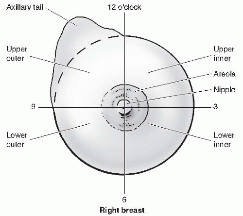
Breast Anatomy Quadrants : The Breasts Amboss : The upper outer quadrant typically contains more fibroglandular tissue than the other quadrants and is where cancers are most likely to develop.. There is a single tumor located at the 12, 3, 6, or 9 o'clock position on the breast code the primary site to c509 when there are multiple tumors (two or more) in at least two quadrants of the breast The upper outer quadrant typically contains more fibroglandular tissue than the other quadrants and is where cancers are most likely to develop. Any preexisting asymmetries, spinal curvature, or chest wall deformities must be recognized and. Breast tissue is drained by lymphatic vessels that lead to axillary nodes (which lie in the axilla) and internal mammary nodes (which lie along each side of the breast bone). 2:00 in the right breast is in the uiq, whereas 2:00 in the left breast is in the uoq.
The fat (subcutaneous adipose tissue) that covers the lobes gives the breast its size and shape. A layer of fatty tissue surrounds the breast glands and extends throughout the breast, which gives the breast a soft consistency and gentle, flowing contour. At the centre of the breast is the nipple, composed mostly of smooth muscle fibres. The breast is a modified sweat gland located in the superficial fascia of the anterior chest wall.the major portion of the breast tissue is situated between the second and third rib superiorly, the sixth and seventh costal cartilage inferiorly, the anterior axillary line laterally, and the sternal border medially. Theory holds that the upper outer quadrant of the breast develops more malignancies because of increased tissue volume.
Breast Diseases Obgyn Key from obgynkey.com The upper outer quadrant extends towards the axilla as the axillary tail. Theory holds that the upper outer quadrant of the breast develops more malignancies because of increased tissue volume. The breast is divided into 4 quadrants. A layer of fatty tissue surrounds the breast glands and extends throughout the breast, which gives the breast a soft consistency and gentle, flowing contour. Each breast contains 15 to 20 lobes arranged in a circular fashion. Embedded in the breast's fatty and fibrous tissue are 15 to 20 glands called lobes, each of which has many smaller lobules, or sacs, that produce milk. Grant's atlas of anatomy, 12th ed., lippincott williams & wilkins. This is because this area has a lot of glandular tissue.
The glandular parenchyma is estrogen dependent, thus on attaining menopause the glandular parenchyma atrophies.
Originates from both the medial and lateral quadrants of the breast passes through the intercostal spaces and pectoralis major into parasternal/internal mammary lymph nodes connections may lead across the median plane and hence to the contralateral breast The four quadrants are upper lateral, upper medial, lower medial, and lower lateral quadrants. Regional lymph nodes blood and lymph vessels form a network throughout each breast. Theory holds that the upper outer quadrant of the breast develops more malignancies because of increased tissue volume. Cancer registration & surveillance modules. A layer of fatty tissue surrounds the breast glands and extends throughout the breast, which gives the breast a soft consistency and gentle, flowing contour. The fat (subcutaneous adipose tissue) that covers the lobes gives the breast its size and shape. The upper outer quadrant extends towards the axilla as the axillary tail. When breast cancer spreads, it is frequently to these nodes. Four quadrants of the breast • upper outer (superolateral) quadrant • upper inner (superomedial) quadrant • lower outer (inferolateral) quadrant. Each lobe is comprised of many lobules, at the end of which are tiny bulb like glands, or sacs, where milk is produced in response to hormonal signals. Lee 11 breast anatomy introduction understanding breast anatomy is important to recognizing the disease processes that may occur within the breast. Nearly all lymphatics of the breast drain along a subdermal plane into the axillae, typically collecting in a single sentinel lymph node at the lateral border of the pectoralis major muscle.
At the centre of the breast is the nipple, composed mostly of smooth muscle fibres. Breast tissue is drained by lymphatic vessels that lead to axillary nodes (which lie in the axilla) and internal mammary nodes (which lie along each side of the breast bone). The breast is divided into four quadrants: The results of this anatomical study may facilitate sn biopsy in patients with breast cancer. There is a single tumor in two or more subsites and the subsite in which the tumor originated is unknown there is a single tumor located at the 12, 3, 6, or 9 o'clock position on the breast code the primary site to c509 when there are multiple tumors (two or more) in at least two quadrants of the breast
Diagnosing Breast Cancer We Don T Just Care We Put In The Effort from i2.wp.com Ducts are thin tubes that carry milk to the nipple. When breast cancer spreads, it is frequently to these nodes. A regional atlas of the human body, 5th ed., lippincott williams & wilkins, baltimore, md, 2006. Theory holds that the upper outer quadrant of the breast develops more malignancies because of increased tissue volume. The fibroglandular tissue is surrounded by mostly fatty tissue in the subcutaneous and retromammary (retroglandular) regions (fig. Anatomy of the breast normal anatomy. The results of this anatomical study may facilitate sn biopsy in patients with breast cancer. The breast can be considered to be composed of two regions:
The glands function is provided by hormones estrogen.
Anatomy of the breast normal anatomy. The glandular parenchyma is estrogen dependent, thus on attaining menopause the glandular parenchyma atrophies. Features of the quadrants of the breast. There is a single tumor in two or more subsites and the subsite in which the tumor originated is unknown there is a single tumor located at the 12, 3, 6, or 9 o'clock position on the breast code the primary site to c509 when there are multiple tumors (two or more) in at least two quadrants of the breast This is because this area has a lot of glandular tissue. The breast is a modified sweat gland located in the superficial fascia of the anterior chest wall.the major portion of the breast tissue is situated between the second and third rib superiorly, the sixth and seventh costal cartilage inferiorly, the anterior axillary line laterally, and the sternal border medially. Cancer registration & surveillance modules. Regional lymph nodes blood and lymph vessels form a network throughout each breast. Each lobe is comprised of many lobules, at the end of which are tiny bulb like glands, or sacs, where milk is produced in response to hormonal signals. Department of health and human services; Embedded in the breast's fatty and fibrous tissue are 15 to 20 glands called lobes, each of which has many smaller lobules, or sacs, that produce milk. Each breast contains 15 to 20 lobes arranged in a circular fashion. Understanding the anatomy of the breast is important in understanding how it.
The upper outer quadrant extends towards the axilla as the axillary tail. Embedded in the breast's fatty and fibrous tissue are 15 to 20 glands called lobes, each of which has many smaller lobules, or sacs, that produce milk. The results of this anatomical study may facilitate sn biopsy in patients with breast cancer. The fibroglandular tissue is surrounded by mostly fatty tissue in the subcutaneous and retromammary (retroglandular) regions (fig. Learn about breast anatomy so you can better understand breast cancer, be aware of anything unusual, & have better dialogue with your doctor.
Female Breast Assessment Flashcards Quizlet from o.quizlet.com Each lobe is comprised of many lobules, at the end of which are tiny bulb like glands, or sacs, where milk is produced in response to hormonal signals. Any preexisting asymmetries, spinal curvature, or chest wall deformities must be recognized and. Each breast contains 15 to 20 lobes arranged in a circular fashion. Learn about breast anatomy so you can better understand breast cancer, be aware of anything unusual, & have better dialogue with your doctor. There is a single tumor in two or more subsites and the subsite in which the tumor originated is unknown there is a single tumor located at the 12, 3, 6, or 9 o'clock position on the breast code the primary site to c509 when there are multiple tumors (two or more) in at least two quadrants of the breast They are localized in the front of the breast within the 3 to 7 rib. Ducts are thin tubes that carry milk to the nipple. A regional atlas of the human body, 5th ed., lippincott williams & wilkins, baltimore, md, 2006.
Lobules are arranged in clusters, like bunches of grapes.
This is because this area has a lot of glandular tissue. The glandular parenchyma is estrogen dependent, thus on attaining menopause the glandular parenchyma atrophies. Ducts are thin tubes that carry milk to the nipple. Originates from both the medial and lateral quadrants of the breast passes through the intercostal spaces and pectoralis major into parasternal/internal mammary lymph nodes connections may lead across the median plane and hence to the contralateral breast Breast shape varies among patients, but knowing and understanding the anatomy of the breast ensures safe surgical planning (see the image below). The breast is divided into 4 quadrants. The breast can be considered to be composed of two regions: Nearly all lymphatics of the breast drain along a subdermal plane into the axillae, typically collecting in a single sentinel lymph node at the lateral border of the pectoralis major muscle. Understanding the anatomy of the breast is important in understanding how it. Features of the quadrants of the breast. The four quadrants are upper lateral, upper medial, lower medial, and lower lateral quadrants. Each breast contains 15 to 20 lobes arranged in a circular fashion. In seven cases (29%), the sn was located in the upper ventral quadrant, in two cases (8%) in the upper dorsal quadrant, and in one case in the lower dorsal quadrant.
A layer of fatty tissue surrounds the breast glands and extends throughout the breast, which gives the breast a soft consistency and gentle, flowing contour anatomy quadrants. Understanding the anatomy of the breast is important in understanding how it.

